Case 3. Numb face
Case
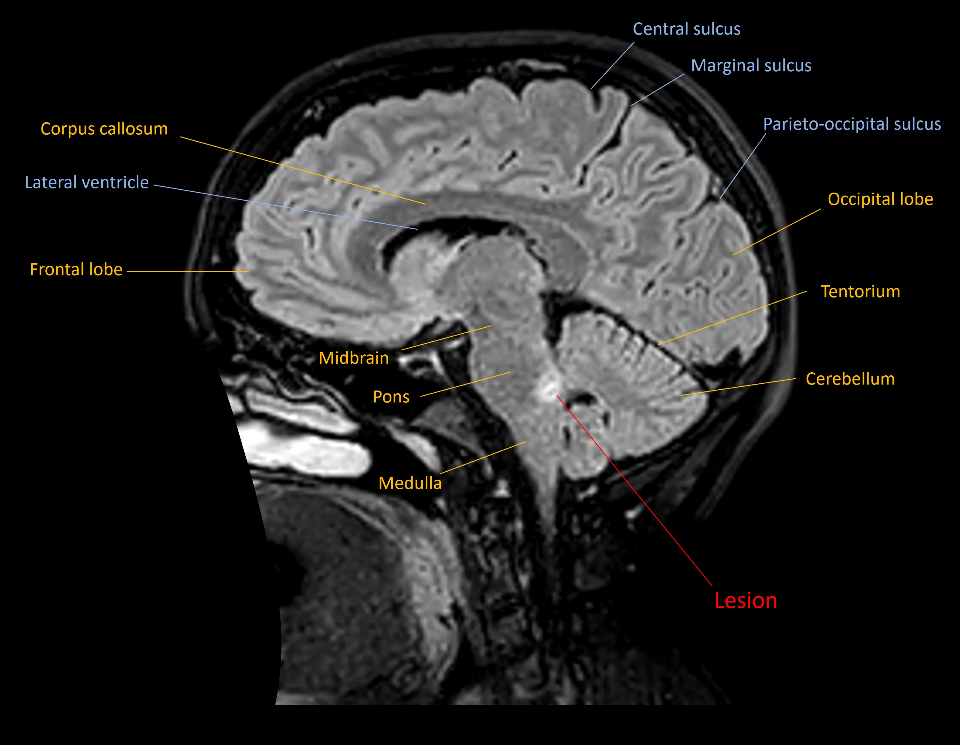
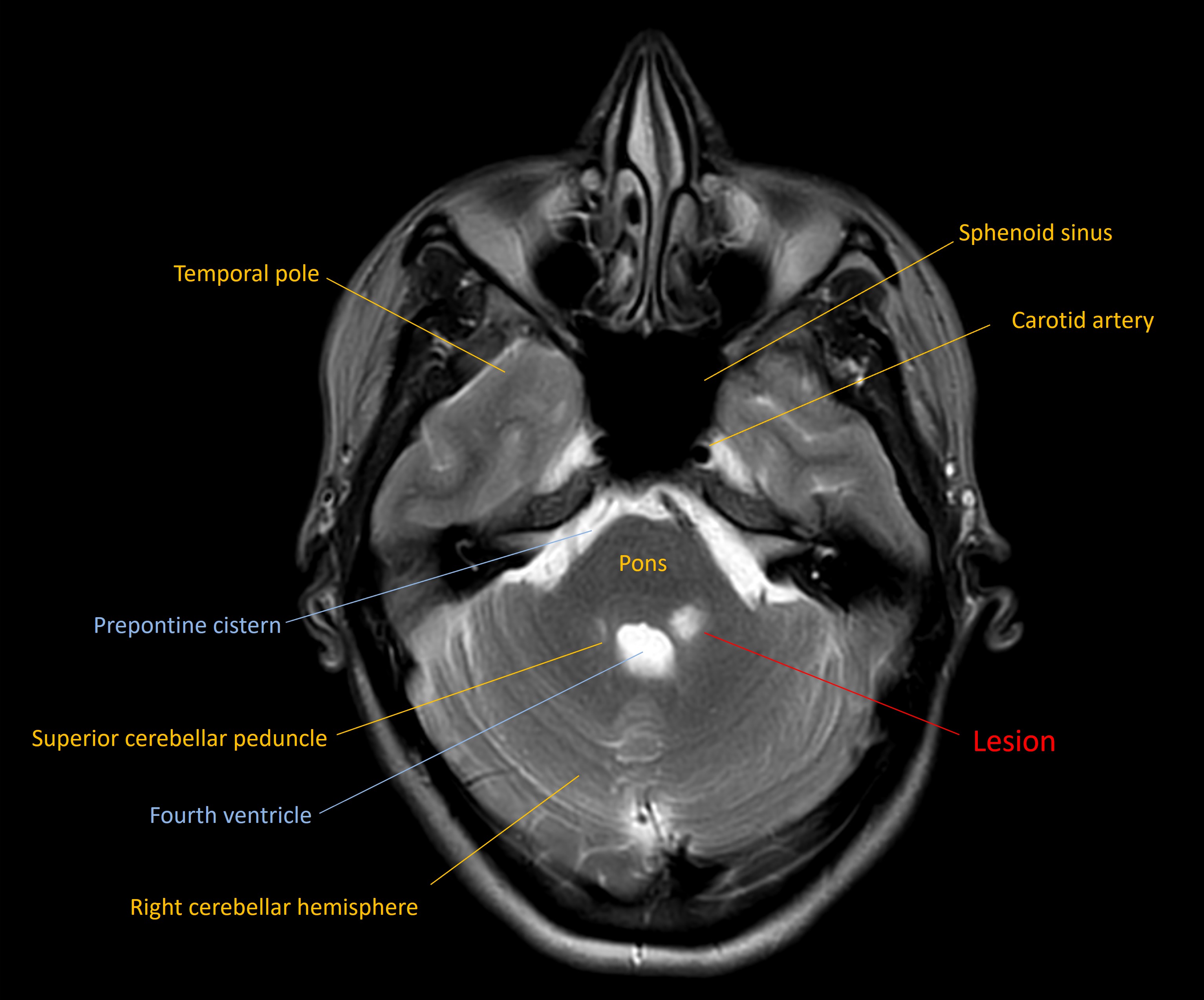
An MRI showed a lesion in the left dorsal upper pons, in the site of the principal trigeminal sensory nucleus - suggestive of demyelination.
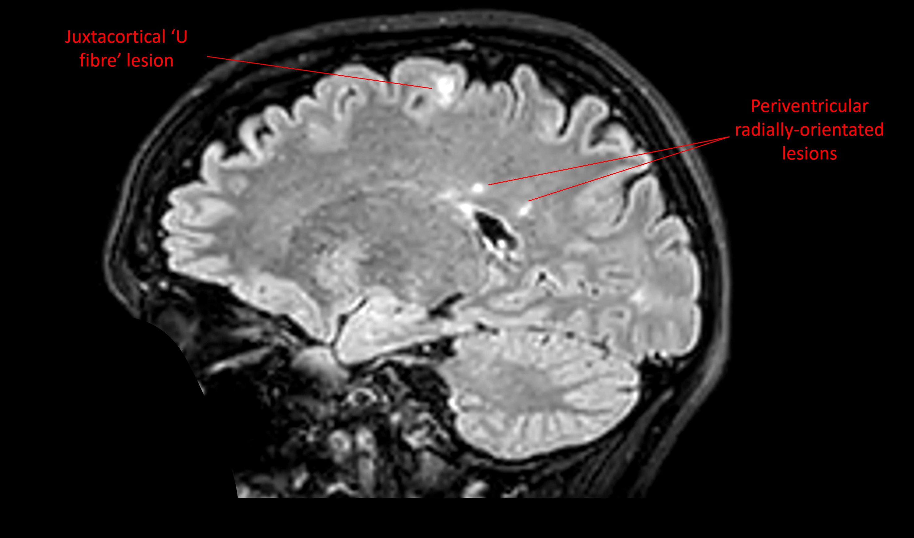
In addition there were multiple white matter lesions in the brain, including a juxtacortical ‘U fibre’ lesion, periventricular lesions, and cerebellar lesions.
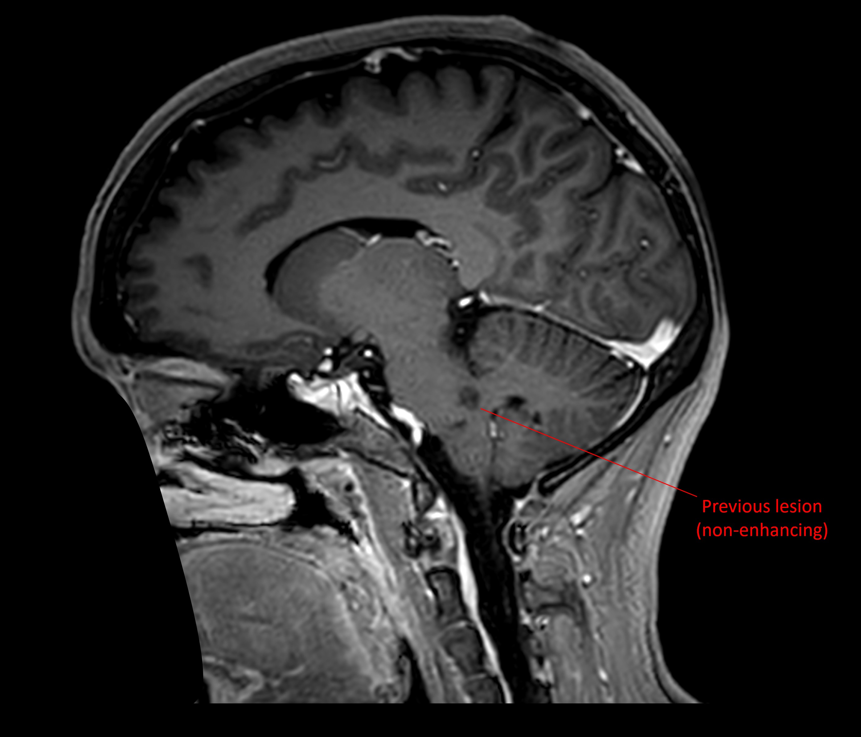
This was suggestive of multiple sclerosis (MS), meeting the criteria for dissemination in space . However, insufficient evidence was present to meet the dissemination in time criteria, so the patient underwent another MRI with contrast 3 weeks later. By this time, the pontine lesion was non-enhancing, while there were new, asymptomatic, enhancing lesions present - demonstrating dissemination in time . MS was diagnosed.
The patient was treated with B-cell depleting infusions, which were tolerated well, with a stable clinical course and no relapses, as well as stable imaging performed annually. The facial sensory loss gradually returned to normal, with only occasional intermittent cheek tingling and numb feelings.
2 years after diagnosis, she paused her infusions for family planning and had an uncomplicated pregnancy, delivering a healthy baby.
Final diagnosisHemifacial numbness due to a demyelinating pontine lesion as the first presentation of MS.
Key pointsReturn to Cases